Testing Plant Substances as Potential Medicines
BACKGROUND: In nature, organisms are constantly battling for resources and survival. Plants compete with other plants for light and water. Fast growth, big leaves, and large root systems would be advantageous characteristics. Some plants, such as rhododendrons, actually produce toxic chemicals that drip from their leaves into the soil, killing competing plants around them.
All organisms are infected by viruses or threatened by bacterial disease. Most organisms have defense systems to protect themselves from potential predators or foreign invaders. Plants such as fungi and bacteria produce antimicrobial agents to battle the microbes. If humans could find and isolate an antimicrobial molecule, it could potentially lead to a potential therapeutic medicine.
Once plant samples are collected, extraction techniques have to be determined. Then, samples can be tested for their ability to kill different types of microbes. Researchers need to prove that certain antimicrobial agents do not cause toxic effects in humans.
To test plant extracts for antimicrobial properties, technicians add extract-soaked filter paper disks to bacteria cultures spread on Petri plates. The extracts demonstrating these clear areas on the Petri dishes are then further purified and screened for the specific ingredients causing the bacterial death. These compounds may include antiseptics, astringents, antibiotics, and toxins. OBJECTIVE/PURPOSE: What plant materials, found locally, contain active ingredients that will inhibit the growth of bacteria?
MATERIALS:-Plant extract
-syringe
-filter paper
-funnel
-microfuge tube
-rack
-pipet
PROCEDURE:Preparing Plant Extracts:
-Using a mortar and pestle, grind up 2 grams of plant tissue with 10 ml of deionized water
-Let it sit for 3 minutes
-Filter the sample through an 11 cm filter paper/funnel.
-Filter sterilize the extract using a syringe filter.
-Collect 1 ml of the filter-sterilized extract into a 1.7 microtube. Label the sample.
Purpose: What plant materials, found locally, contain active ingredients that will inhibit the growth of bacteria?
Procedure:
Analysis and Observations:
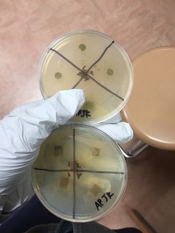
Day 1: We looked at our petri dishes to see if the bacteria grew and showed the results; the results being whether there was a clearance around our paper disks. We didn't see too much of a difference, and you could barely tell whether there was a clearance or not.
Day 2: We saw a small difference from day one. It was hard to decipher the difference between the cloudy bacteria and the clear agar. There was a small clearance around the disks, but like said above, it was hard to tell.
BACKGROUND: In nature, organisms are constantly battling for resources and survival. Plants compete with other plants for light and water. Fast growth, big leaves, and large root systems would be advantageous characteristics. Some plants, such as rhododendrons, actually produce toxic chemicals that drip from their leaves into the soil, killing competing plants around them.
All organisms are infected by viruses or threatened by bacterial disease. Most organisms have defense systems to protect themselves from potential predators or foreign invaders. Plants such as fungi and bacteria produce antimicrobial agents to battle the microbes. If humans could find and isolate an antimicrobial molecule, it could potentially lead to a potential therapeutic medicine.
Once plant samples are collected, extraction techniques have to be determined. Then, samples can be tested for their ability to kill different types of microbes. Researchers need to prove that certain antimicrobial agents do not cause toxic effects in humans.
To test plant extracts for antimicrobial properties, technicians add extract-soaked filter paper disks to bacteria cultures spread on Petri plates. The extracts demonstrating these clear areas on the Petri dishes are then further purified and screened for the specific ingredients causing the bacterial death. These compounds may include antiseptics, astringents, antibiotics, and toxins. OBJECTIVE/PURPOSE: What plant materials, found locally, contain active ingredients that will inhibit the growth of bacteria?
MATERIALS:-Plant extract
-syringe
-filter paper
-funnel
-microfuge tube
-rack
-pipet
PROCEDURE:Preparing Plant Extracts:
-Using a mortar and pestle, grind up 2 grams of plant tissue with 10 ml of deionized water
-Let it sit for 3 minutes
-Filter the sample through an 11 cm filter paper/funnel.
-Filter sterilize the extract using a syringe filter.
-Collect 1 ml of the filter-sterilized extract into a 1.7 microtube. Label the sample.
Purpose: What plant materials, found locally, contain active ingredients that will inhibit the growth of bacteria?
Procedure:
- Preparing plant extracts:
- Use a mortar and paste to grind up 2 grams of plant tissue with 10 ml of deionized water
- Let it sit for 3 minutes
- Filter the sample through an 11 cm filter paper/funnel
- Filter/sterilize the extract using a syringe filter
- Collect 1 mL of the filter-standard extract into a 17 microtube. Label the sample
- Attach prefilter to syringe and rinse with water
- Take to Laminar hood: Plant extract, Syringe/prefilter, Microfuge tube rack, Pipet
- Label microfuge tube with initials and whether it is water/methanol and put in rack
- Attach the sterile filter to the prefilter
- Load 1.7 mL of extract to syringe using pipet
- Put plunger in and depress it
- Have at least 1 mL of sterilized extract
- Snap on cap on microfuge tube
- Evaporate methanol from methanol extracts by placing a tube, with a cap, upon a 65 degree heat-block overnight
- Reconstitute methanol extract 1 mL sterile deionized water
- Using sterile forceps place 3 sterile pieces of filter paper into the filtered extract 4 degree celsius
- Store until ready to use
- Draw a + on each plate bottom and number quadrants 1-4
- Liquify sterile LB agar in the microwave
- Using sterile technique pour approximately 20 mL of agar into Petri plate
- Using sterile forceps add the appropriate number of sterile disks to each tube of filtered extract
- Label both plates with either M for methanol or W for water
- Place the disks into the appropriate solution
- Sterile disks were added to microfuge tubes containing one ML sterile water
- 10-20 ml of warmed nutrient agar was poured into 2 petri dishes wsing steik techniques
- After allowing agar to solidify, plates were turned upside down and stored at 4 degrees celsius overnight
- One ml of E.coli colony was added to each plate. A flame-sterilized spreading loop was used to spread the bacteria throughout the surface of the agar.
- Using flame-sterilized forceps, filter desks were placed in separate quadrants onto the plate in the following sequence: 1) water 2) plant extracts 3) ampicillin. Plates were left on lab bench for 20 min. to allow both bacteria and filter disks to adhere to the agar.
- plates were incubated upside down, overnight at 37 degreesC. Plates were photographed and observed for clearance around the filter disks after 24 hours, 48 hours, 72 hours
Analysis and Observations:
Day 1: We looked at our petri dishes to see if the bacteria grew and showed the results; the results being whether there was a clearance around our paper disks. We didn't see too much of a difference, and you could barely tell whether there was a clearance or not.
Day 2: We saw a small difference from day one. It was hard to decipher the difference between the cloudy bacteria and the clear agar. There was a small clearance around the disks, but like said above, it was hard to tell.